If you’re interested in dermatology, your rotation is the perfect opportunity to explore the specialty, build hands-on experience, and see if it’s the right path for you. Whether you’re hoping to pursue dermatology after graduation or just want to make the most of your rotation, these tips will help you make a learning opportunity out of every case you see.
To help you get the most value from this experience, we spoke with Audrey, a board-certified dermatology PA and advocate for inclusive skincare and medical education. Her highly thoughtful, firsthand insights and practical tips throughout this blog will help you approach your rotation with curiosity, confidence, and a strong clinical foundation.
Review Common Dermatologic Conditions
Before your rotation begins, you’ll benefit from building strong foundational knowledge. Prioritize reviewing the conditions you’ll see most often in outpatient clinics. Start by identifying the highest-yield diagnoses and understanding their clinical features, treatments, and visual presentations.
During my rotation, I made a list of diagnoses I saw repeatedly in the clinic and reviewed those first. It helped me stay one step ahead.”
Your PA QBank is an asset that can help you begin this process. Log into your account and select “dermatologic” under the “systems” category to review relevant questions and reinforce key conditions before encountering them in real life.
To help you get started, we’ve outlined the most common dermatologic conditions below, along with key features and treatments you’ll want to know before your rotation begins.
| Condition | Typical Symptoms | Common Treatments |
| Acne (Acne Vulgaris) | Blackheads, whiteheads, pimples, cysts | Topical/oral retinoids, antibiotics |
| Eczema (Atopic Dermatitis) | Dry, itchy, inflamed skin; may flare with triggers | Moisturizers, topical steroids, immunomodulators |
| Psoriasis | Thick plaques with silvery scales, especially on elbows/knees/scalp | Topicals, phototherapy, systemic treatments (e.g., biologics) |
| Seborrheic Dermatitis | Yellow/white scaly patches on scalp, face | Medicated shampoos, antifungal creams |
| Rosacea | Facial redness, visible vessels, pustules; triggered by heat/spicy foods | Topical metronidazole, oral antibiotics, laser therapy |
| Hives (Urticaria) | Itchy, raised welts that come and go | Antihistamines, corticosteroids |
| Fungal Infections | Itchy, scaly rashes (e.g., ringworm, athlete’s foot) | Topical/oral antifungals |
| Basal Cell Carcinoma | Shiny or scaly lesion on sun-exposed skin | Surgical excision, topical chemotherapy, radiation |
| Contact Dermatitis | Redness, itching after contact with irritants or allergens | Trigger avoidance, topical corticosteroids |
| Melasma | Dark facial patches, often linked to hormones or sun exposure | Sun protection, topical depigmenting agents |
Source: 1
Learn Dermatologic Terminology
Not only is it helpful to go into your rotation with diagnostic knowledge, it is equally important to learn the language of the field.
Speaking like a clinician means describing what you see in a way your preceptor understands, beginning with adopting dermatologic terminology. Being able to communicate your observations using the correct terminology not only shows professionalism, but also helps you think more critically about what you're seeing.
Describing a rash using morphology, size, color, and distribution helps your preceptor picture it before even seeing the patient.”
Reviewing key terms ahead of time can help you follow clinic conversations and contribute to assessments with confidence. That said, you’re not expected to know everything, so don’t hesitate to ask questions or clarify unfamiliar terms as you go. Your rotation is a learning experience, and curiosity is always a positive thing.
Describe the primary type of lesion that has arisen spontaneously and reflects the disease process — for example, a macule, papule, plaque, vesicle, or pustule.
Include lesion size using standard measurements; for example, macules are < 1 cm, while patches are > 1 cm; papules are < 1 cm in height, and plaques are > 1 cm.
Note whether the lesion has well-defined or poorly defined borders, which can help distinguish between conditions like tinea corporis (well-demarcated) and eczema.
Use accurate dermatologic color terms such as erythematous (red), hyperpigmented (darkened), hypopigmented (lightened), depigmented (complete color loss), pink, or dusky.
Describe changes from trauma or progression of a primary lesion, for example, scaling in psoriasis or crusting in impetigo.
Comment on the lesion’s pattern and location, such as localized vs. generalized, symmetrical vs. asymmetrical, or specific patterns like dermatomal (zoster) or intertriginous (candidiasis).
Source: 2


Understand Dermatologic Procedures
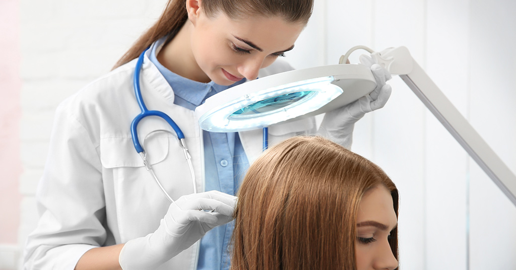
Dermatology is a hands-on specialty, and you’ll likely assist with procedures during most patient visits. From biopsies to cryotherapy, these techniques are essential to patient care, and your rotation is the perfect place to familiarize yourself with them.
One of the best things you can do is have your gloves on and start setting up for the procedure as soon as you hear what’s coming.”
You don’t need to be an expert, but understanding the different types of biopsies and why they are used can help you follow your preceptor’s clinical reasoning.
This technique is often used for superficial lesions, such as seborrheic keratoses or suspected basal cell carcinoma.
A punch biopsy is useful for sampling deeper lesions or inflammatory rashes that require histologic diagnosis.
This procedure is typically reserved for larger growths or pigmented lesions suspicious for melanoma, where complete removal is necessary.
Cryotherapy involves freezing lesions with liquid nitrogen and is commonly used to treat actinic keratoses, warts, or benign lesions.
Curettage is a scraping technique often paired with electrodessication, commonly used to treat small, superficial skin cancers like superficial basal cell carcinoma.
This tissue-sparing technique is used to treat basal and squamous cell carcinomas, especially on the face and other sensitive areas.
Source: 3
Beyond Preparation: Tips for Your Time On-Site
Preparation doesn’t end on day 1 of your dermatology rotation. In fact, some of the most valuable learning happens while you’re in the clinic.
Once you’re on-site, there are still plenty of ways to stay engaged, continue learning, and contribute meaningfully to patient care. From anticipating procedures to supporting the team, these strategies will help you turn everyday moments into opportunities for growth.
If your preceptor is looking at slides for fungus, yeast, or scabies, ask to take a look, too. You’ll learn so much just by being curious.”
Here are a few additional tips to maximize your experience:
- Set up for success: Take initiative by learning how to prepare trays for common procedures like shave, punch, or excisional biopsies. Anticipating what your preceptor needs not only keeps the clinic running smoothly but also shows you're paying attention.
- Stay patient-focused: Simple actions like helping patients get dressed, jotting down post-visit instructions, or clarifying prescription routines (morning vs. evening) can go a long way in supporting both the patient and the clinical team.
- Be curious and ask questions: Whether it's about biopsy results, procedural steps, or treatment decisions, thoughtful questions can deepen your understanding and show your commitment to learning. Don’t be afraid to speak up — your rotation is the time to ask.
- Practice professionalism: Always be mindful of patient comfort, especially during skin exams. Avoid standing too close or hovering while patients are undressed, and maintain a respectful, reassuring presence.
- Prepare ahead of time: If you’re able to view the clinic schedule in advance, take time to look up unfamiliar diagnoses or treatments. This helps you come in with context and allows you to contribute more meaningfully to discussions.
- Support the team: Pitch in wherever you can. Tidy up exam rooms, restock supplies, or offer to help with documentation when appropriate. These small tasks show teamwork and make a strong impression.
Above all, stay approachable and open to learning. Building rapport with staff, preceptors, and patients creates a better environment for everyone and sets the tone for a successful rotation.
Go-To Resources for Dermatology Rotations
Having quick, reliable resources on hand can make your dermatology rotation smoother and more educational. Whether you’re looking up conditions between patients or reviewing biopsy techniques after clinic, the right tools can help reinforce what you’re learning in real time.
VisualDx was my favorite app to use in dermatology. It shows concise details about conditions and images across a range of skin types. A must!”
In addition, many institutions offer free access to clinical apps and digital textbooks that can support your learning throughout the rotation. Here are a few trusted resources to consider:
- Fitzpatrick’s Color Atlas and Synopsis of Clinical Dermatology: A visual-heavy reference with concise summaries — great when you need to quickly compare images of common conditions.
- Habif’s Clinical Dermatology: An accessible, student-friendly introduction to dermatologic diagnoses and treatment plans.
- Lookingbill and Marks’ Principles of Dermatology: Similar in style and scope to Habif, with clear explanations and strong visuals for quick reference.
- VisualDx: Offers brief overviews and photos of conditions across a wide range of skin tones.
- UWorld Mobile App: Provides expertly written questions, visual explanations, flashcards, and performance tracking synced across devices.
- Epocrates: Useful for checking medication dosages, interactions, and prescribing guidelines on the go.
- UpToDate: Offers evidence-based summaries, treatment algorithms, and clinical trial data to support decision-making.
- UWorld PA Medical Library: Provides answers to clinical questions with expert-reviewed articles, high-yield visuals, and integrated tools for deeper learning.
References
- American Academy of Dermatology. (n.d.). Stats and facts.https://www.aad.org/media/stats-numbers
- Stanford Medicine 25. (n.d.). Dermatologic exam. Stanford University School of Medicine. https://stanfordmedicine25.stanford.edu/the25/dermatology.html
- American Osteopathic College of Dermatology. (n.d.). Dermatologic procedures.https://www.aocd.org/page/DermProcedures